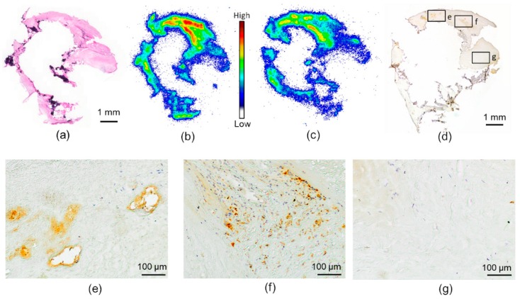
Figure 2.
[18F]Flutemetamol in vitro autoradiography of human carotid atherosclerotic plaque. (a) Hematoxylin-eosin-stained section of human carotid endarterectomy sample. Calcified areas are seen as dark purple; the rest of the plaque represents fibroatheroma. (b) [18F]Flutemetamol in vitro autoradiography of the same section as in A. High uptake is seen as red in specific areas of the plaque. (c) In vitro autoradiography with an excess amount of unlabeled Pittsburgh compound B (PIB) shows decreased accumulation of [18F]Flutemetamol in the plaque. The same scale as in (b). (d) Aβ staining of a consecutive section of the carotid artery plaque. (e–g) Magnification of the areas specified in (d). Aβ-positivity is observed in the areas with high [18F]Flutemetamol accumulation, whereas no staining is seen in the area of low accumulation.

