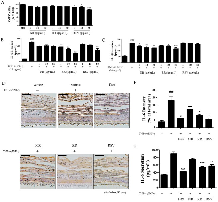
Figure 4.
Inhibitory effects of RR on inflammatory cytokine expression in TNF-α/INF-γ-induced HaCaT cells and 3D skin tissue. (A) Cells were cultured in 96-well plates and treated with NR, RR, and RSV (1, 10, and 50 μg/mL, respectively). After 24 h, cell viability was measured using the MTT assay. (B, C) Cells were treated with NR, RR, and RSV in the presence of TNF-α/IFN-γ (each 10 ng/mL). After 24 h, IL-6 and IL-1β levels in supernatants were determined by ELISA. (D) The expression of IL-6 in TNF-α/IFN-γ-induced 3D skin model was evaluated by IHC. Vehicle (polyethylene glycol: PBS, 1:1), Dex (1%), NR (1%), RR (1%), and RSV (1%) were applied after TNF-α/IFN-γ treatment in 3D skin model thrice weekly. (E) Graphical representation of IL-6 expression in epidermal tissue. Scale bar, 50 μm. (F) The level of IL-6 secretion in media was measured using ELISA. Values are expressed as means ± SEM. ### p < 0.001 versus control group; * p < 0.05, ** p < 0.01, and *** p < 0.001 versus TNF-α/IFN-γ-treated group.

