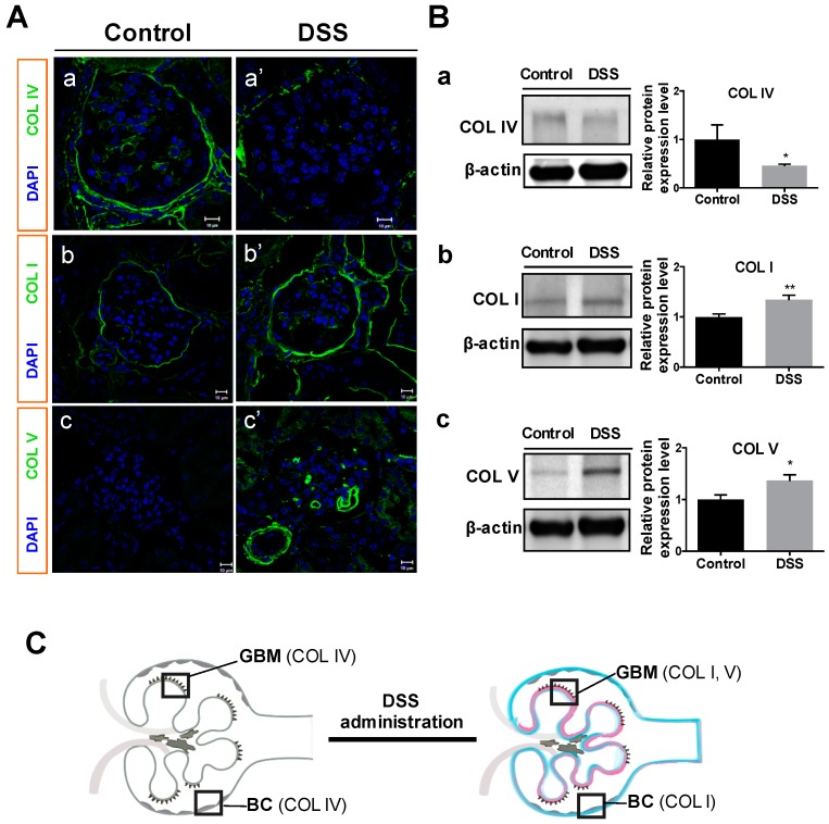
Figure 4.
Changes in glomerular collagens in mice after DSS administration. Immunofluorescent microscopy (A) and Western blot analysis of protein expression (B) for type IV collagen (COL IV; A-a, A-a’; B-a; 160–190 kDa), type I collagen (COL I; A-b, A-b’; B-b; 150 kDa), and type V collagen (COL V; A-c, A-c’; B-c; 220 kDa) were conducted for control and DSS-colitis mice. Representative bands (B, left) and relative band intensity ratios were analyzed (B, right). (C) Illustration of glomerular collagens changes in this study. All values are means ± SEM (n = 6); * p < 0.05 and ** p < 0.01 vs. control. Scale bars = 10 μm. Abbreviations: GBM, glomerular basement membrane; BC, Bowman’s capsule.

