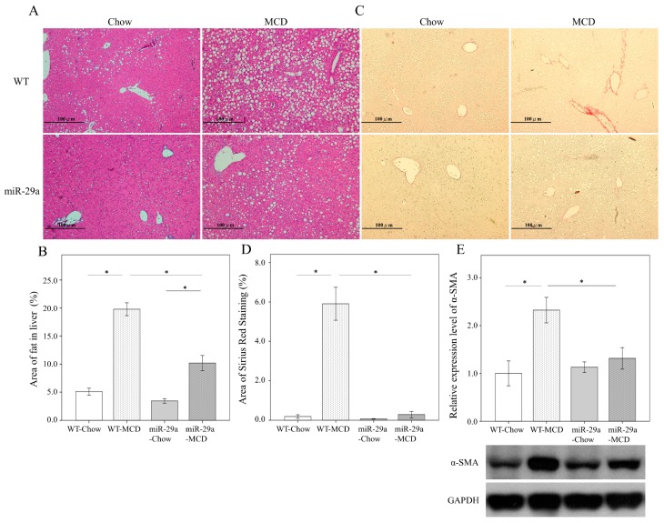
Figure 1.
Overexpression of miR-29a significantly reduced hepatocellular steatosis and fibrosis in methionine-choline-deficient (MCD) diet mice. The hematoxylin and eosin stain showed an abundance of fat droplets accumulated in the liver after feeding WT mice the MCD diet for 4 weeks (A,B, p < 0.001). Compared to the WT littermates, miR-29a overexpression reduced the abundance of fat droplets accumulated in the livers of miR-29aTg mice (A,B, p < 0.001). Furthermore, Sirius red stain and western blot demonstrated a greater accumulation of extracellular matrix (C,D) and α-SMA expression (E) in the livers of WT-MCD mice compared to WT-Chou (both p < 0.001). However, miR-29a-MCD mice showed a weaker induction of collagenous matrices and α-SMA compared to WT-MCD mice (p < 0.001 and p = 0.024, respectively). Data are expressed as the mean ± SD of six to eight samples per group. * indicates a p < 0.05 between the groups.

