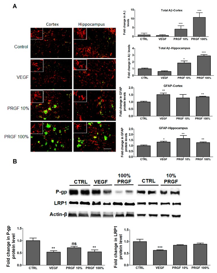
Figure 5.
Effect of VEGF (100 pg/mL) and PRGF (10 and 100%) on Aβ burden and astrocytes activation in the hippocampus and cortex of 5xFAD mice. (A) Representative hippocampus and cortex sections and optical density quantification of Aβ and GFAP in 5xFAD mice treated with VEGF and PRGF; sections were stained with 6E10 antibody against Aβ to detect total Aβ load (green), and anti-GFAP antibody (red) to detect activated astrocytes. The hippocampus images were from the CA1 region; however, the entire hippocampus region was included in the quantification spanning the dentate gyrus and CA1-CA3 regions. Images in white boxes are showing astrocytes at higher magnification. (B) Representative blots and densitometry analysis of P-gp and LRP1 in microvessels isolated from 5xFAD mice brains. Data are presented as mean ± SEM (n = 7 mice/group). * p < 0.05, ** p < 0.01, *** p < 0.001 compared to control (CTRL) group. The white scale bar indicates 50 µm length.

