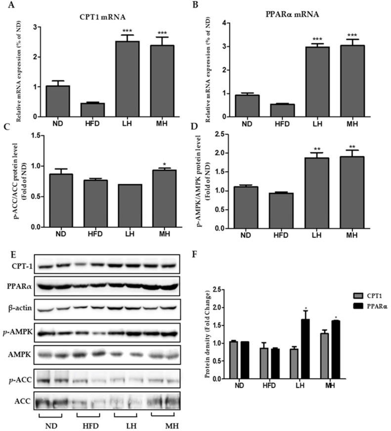
Figure 8.
Effects of HBE on CPT-1 and PPARα expression and AMPK, ACC signaling in HFD-fed mice. The expression of CPT-1 (A) and PPARα (B) were quantified by real-time PCR and normalized by β-actin as an internal control. (C–D) Western blot analysis of p-AMPK/AMPK and p-ACC/ACC protein in the livers of mice fed an ND, HFD, or HFD with supplemented HBE (LH, 0.5% or MH, 1%). β-actin was used as a protein loading control. AMPK and ACC were used as protein loading controls of phosphorylated AMPK (p-AMPK) and phosphorylated ACC (p-ACC), respectively. (E) CPT-1, PPARα, phosphorylated AMPK, and phosphorylated ACC protein levels by immunoblot analysis. (F) Protein density of CPT-1, PPARα. Equal loading of protein was verified by probing β-actin. Data represent means ± SD. * p < 0.5, ** p < 0.01, *** p < 0.001 vs. HFD.

