Figure 2.
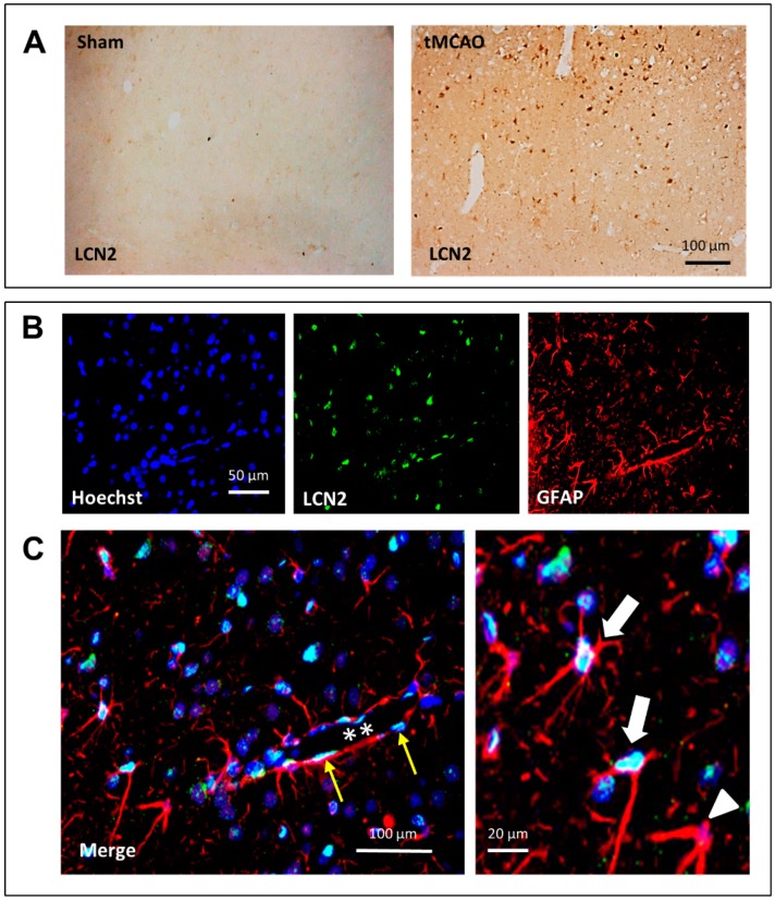
Immunohistochemical (A) and double-immunofluorescence (B) staining of LCN2 after stroke in the cerebral cortex of tMCAO rats. (A) Numbers of LCN2-positive cells massively increased after tMCAO. In sham-operated animals, almost no LCN2-positive cells were visible. (C) Double-labeling revealed that many GFAP-positive (red) astrocytes are also positive for LCN2 (turquoise-white). In (C), a blood vessel (stars) crosses the section, and thin elongated endothelial cells (yellow arrows) can be identified. At higher magnification, two LCN2-positive (white arrows) and a LCN2-negative (white arrowhead) can be seen.

