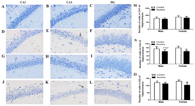
Figure 2.
Morphological evaluation of Nissl staining in CA1, CA3 and DG regions of the hippocampus in adolescent rats. (A-C) Morphology of the hippocampus in male control group (magnification, ×40). (D-F) Morphology of the hippocampus in male PNE (magnification, ×40). (G-I) Morphology of the hippocampus in female control group (magnification, ×40). (J-L) Morphology of the hippocampus in female PNE (magnification, ×40). Arrows indicate the dissolved Nissl bodies. (M-O) The number of cells in CA1, CA3 and DG of rat hippocampus. Five sections of each group were selected and five random fields of each section were scored. The data are expressed as mean ± standard error of the mean; n=4 offsprings; *P<0.05, **P<0.01 vs. control. PNE, prenatal nicotine exposure.

