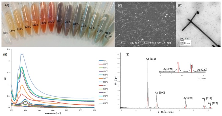
Figure 1.
Characterization of silver nanowires-AgNWs: (A) Typical color change of the reaction mixture at different temperatures; (B) the corresponding Ultra Violet spectra; (C) Scanning Electron Microscopy image of AgNWs suspended in a solution of chitosan in lactic acid (scale bar 4 µm); (D) Transmission Electron Microscopy image of AgNWs (scale bar 100 nm); (E) X Ray Diffraction pattern of the synthesized AgNWs (red bars: Ag, PDF No. 04-0783; blue bars: AgCl, PDF No. 31-1238).

