
Figure 7. Macrophage Mitochondrial Dysfunction Impairs IL-10-Associated Cardiac Repair.
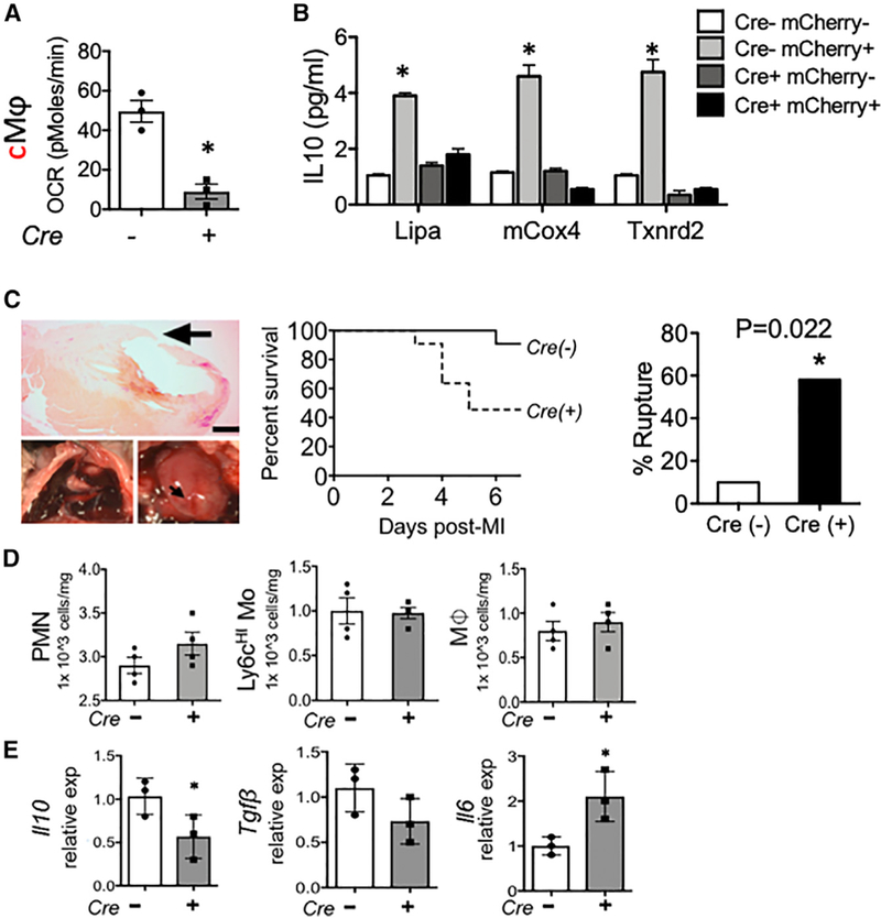
Rispfl/fl and Rispfl/fl LysMcre mice were subjected to experimental myocardial infarction (MI) at the left anterior descending artery, as described in STAR Methods. (A) Cardiac extracts were prepared and flow cytometry employed to isolate left ventricular cardiac Mᶲs (CD45+CD11b+F4/80+Ly6g—CD64+). Mice transgenic for cardiac-specific expression of the fluor mCherry were subjected to MI, and cardiac Mᶲs containing mCherry were interrogated for OCR in Rispfl/fl LysMcre mice. (B) Gene expression by qPCR from indicated cell types, sorted directly from heart. (C) Representative images and Kaplan-Meier survival plot and rupture quantification of indicated mice after MI. n = 10 (Cre—) versus 11 (Cre+) and p = 0.02. (D) Levels of indicated immune cell subsets (PMN, neutrophil; Mo, monocyte; Mᶲ, macrophage) after MI. (E) Gene expression of indicated inflammatory mediators from cardiac extracts. *p < 0.05.