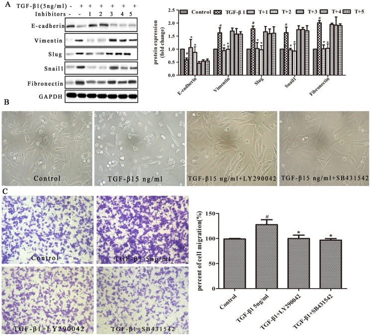
Figure 4.
TGF-β1 induced EMT in MDA231 cells through the PI3K/AKT and Smad pathways. (A) Cells were treated with or without the following for 24 h: 5 ng/mL TGF-β1; 1, LY290042 (PI3K inhibitor); 2, SB431542 (ALK4/5/7 inhibitor); 3, SP600125 (JNK inhibitor); 4, PDTC (NF-κB inhibitor); 5, PD98059 (ERK inhibitor). The proteins were evaluated by Western blot analysis. The values are expressed as the mean ± SD. The experiments were repeated three times. # p < 0.05 vs. untreated cells; * p < 0.05 vs. cells treated with 5 ng/mL TGF-β1. (B) Morphology of MDA231 cells treated with TGF-β1 and inhibitors. The images were captured by brightfield microscopy (200× magnification). Cells were treated with or without 5 ng/mL TGF-β1, TGF-β1+LY290042 or TGF-β1+SB431542 for 24 h. TGF-β1 induced morphological changes in mesenchymal cells in the MDA231 cell line: intercellular connections disappeared. However, this effect was reversed by the inhibitors. (C) Transwell chambers were used to detect the extent of cell migration (×100 magnification). MDA231 cells were untreated or treated with 5 ng/mL TGF-β1, 10 μM LY290042 and 5 ng/mL TGF-β1, or 10 μM SB431542 and 5 ng/mL TGF-β1 for 24 h. The percent cell migration is shown. The error bars represent three independent experiments, and each experiment was repeated three times. * p < 0.05 vs. cells treated with 5 ng/mL TGF-β1. # p < 0.05 vs. untreated cells.

