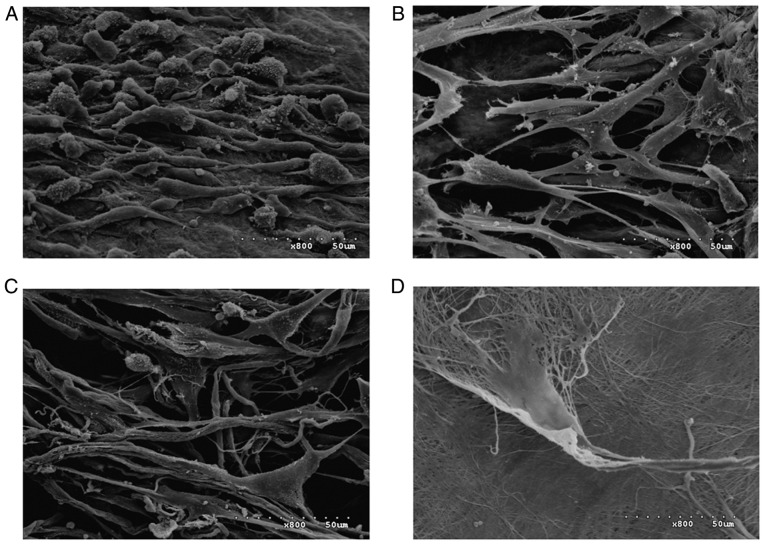
Figure 11.
Scanning electron microscopy images of the four scaffolds seeded with HGFs and cultured for 7 days (magnification, ×800). (A) Cells-ACVM-0.25% HLC-I, (B) Cells-ADM, (C) Cells-Bio-Gide and (D) Cells-SIS. A large number of HGFs were visible on the surface of the ACVM-0.25% HLC-I scaffold. The SIS scaffold had the smallest number of HGFs on the surface. ACVM-0.25% HLC-I, acellular vascular matrix-0.25% human-like collagen I; ADM, acellular dermal matrix; SIS, small intestinal submucosa; HGF, human gingival fibroblast.

