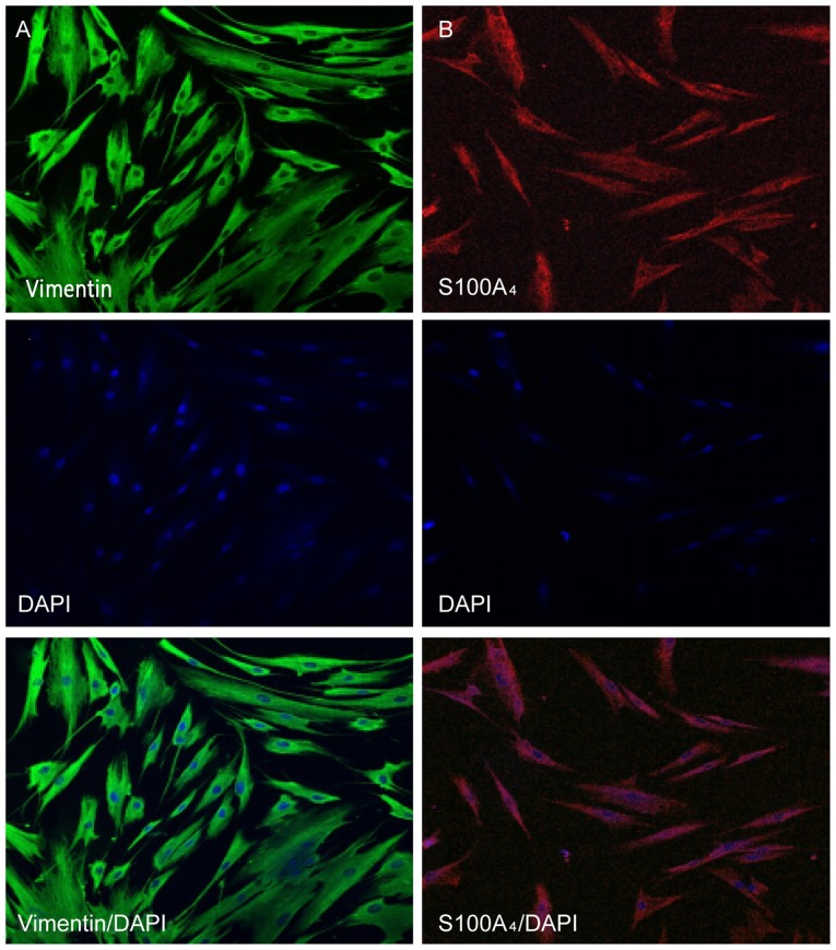
Figure 3.
Immunofluorescent staining of HGFs. (A) Cytoplasm of HGFs with positive staining for vimentin (fluorescein isothiocyanate; magnification, ×200). (B) Cytoplasm of HGFs with positively stained for S100A4 (Rhodamine; magnification, ×200). Vimentin and S100A4 are markers for mesenchymal cells. DAPI is a marker of cell nuclei. HGF, human gingival fibroblast; DAPI, 4′,6-diamidino-2-phenylindole.

