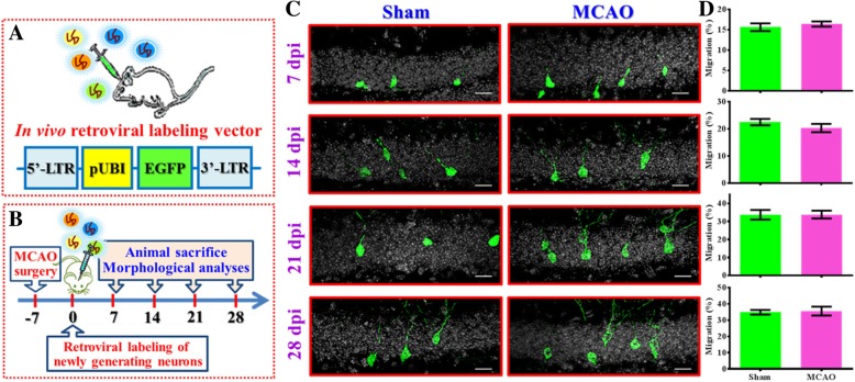
Fig. 1.
Retroviral labeling of newly generated neurons after stroke. (a) The retroviral vector used in the study for labeling and tracing the newly generated neurons after MCAO surgery in vivo. LTR: Long terminal repeats, pUBI: Ubiquitin promoter, EGFP: enhanced green fluorescent protein. (b) Schematic illustration of the procedure of experimental designs. (c) Representative images of the retroviral labeled newly generated neurons obtained at 7, 14, 21 or 28 days-post viral-injection (dpi). Green, GFP signal from labeled newborn neurons. White, DAPI signal for the granular cell layer. The scale bar is 25 μm. (d) Quantification of the relative migratory distribution of the soma from the labeled cells along the granular cell layer at 7, 14, 21 or 28 dpi. Data are presented as mean ± SEM. n = 30 to 50 cells from four mice, two-tailed unpaired t-test

