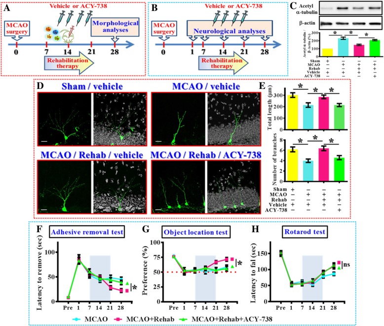
Fig. 8.
Requirement of HDAC6 in rehabilitation therapy induced functional recovery. (a) Schematic illustration the procedure for studying HDAC6 in rehabilitation therapy induced reversal of dendritic phenotypes. (b) Schematic illustration the procedure for studying HDAC6 in rehabilitation therapy induced long term functional recovery after MCAO. (c) Representative western blot images and quantification of the acetylated-α-tubulin that normalized to β-actin from different experimental groups. n = 5 biological replicates, *p < 0.05. (d) Representative images of the newly generated neurons for their dendritic elaboration at 14 dpi. “Rehab” indicates the rehabilitation therapy for the mice. The scale bar is 25 μm. (e) Quantification of total dendritic length and the number of dendritic branches of individual newborn neurons at 14 dpi from different groups. Data are presented as mean ± SEM. n = 20 to 40 cells from four mice, one-way ANOVA with Bonferroni’s post hoc analysis. * p < 0.05. (f) Behavioral performance of adhesive removal task. Two-way ANOVA analysis showed significant difference between the treatments (p < 0.001) and Bonferroni’s post hoc: p < 0.05 for the time points of 28 days for the MCAO+Rehab vs. MCAO+Rehab+ACY-738. (g) Object location memory test before or days after MCAO. Two-way ANOVA analysis showed significance between the treatments (p < 0.01) and Bonferroni’s post hoc: p < 0.05 for the time points of 28 days for the MCAO+Rehab vs. MCAO+Rehab+ACY-738. (h) Rotarod test before or days after MCAO. Two-way ANOVA analysis showed significant difference between the treatments (p < 0.01) and Bonferroni’s post hoc showed no significance for the time points of 28 days for the MCAO+Rehab vs. MCAO+Rehab+ACY-738. n = 10 mice

