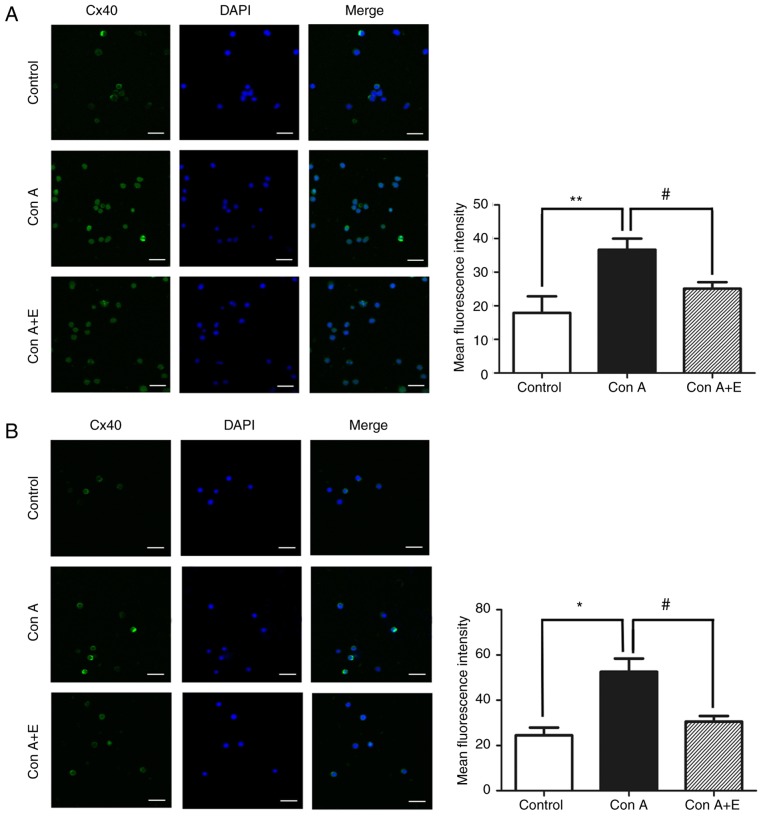
Figure 8.
β-estradiol pre-treatment decreased Cx40/Cx43 expression in peripheral blood lymphocytes stimulated with Con A. Representative immunofluorescence images of (A) Cx40 and (B) Cx43 (magnification, ×630; scale bar, 16 µm) to determine their localization and expression in the plasma membrane and cytosol. These images presenting the changes of Cx40 and Cx43 expression when Con A stimulated peripheral blood lymphocytes were pretreated with β-estradiol. Bar graphs presenting densitometric analysis of mean immunofluorescent intensity of Cx40 and Cx43 in various groups. Each figure is representative of three independent experiments. Each column represents the mean ± standard error from five samples/group. *P<0.05 or **P<0.01, control group vs. Con A-stimulated group; #P<0.05, Con A-stimulated group vs. E-treated group. Cx, connexin; Con A, concanavalin A; DAPI, 4′,6-diamidino-2-phenylindole; E, β-estradiol.

