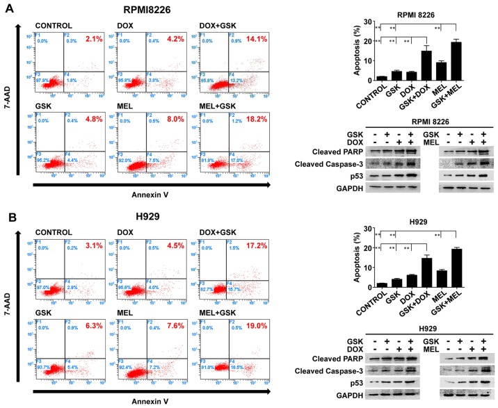
Figure 3.
GSK 126 augments DNA-damaging agent-induced apoptosis in MM cells. (A) RPMI8226 and (B) H929 cells were treated with GSK126 (5 µM), DOX (0.1 µM), MEL (10 µM) or a combination treatment for 48 h. The percentage of apoptotic cells was established by flow cytometry with Annexin V-phycoerythrin and 7-aminoactinomycin D staining. **P<0.01. Whole-cell lysates were utilized to evaluate the protein expression of cleaved PARP, cleaved caspase-3 and p53 via western blotting. GAPDH was utilized as the loading control. All the experiments represent the mean ± standard deviation from three independent experiments. DOX, doxorubicin; MEL, melphalan; PARP, poly (ADP-ribose) polymerase; p53, cellular tumor antigen p53.

