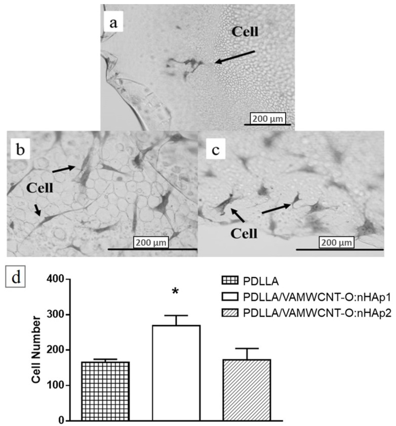
Figure 3.
Optical microscopy of the human chondrocytes adhered on (a) PDLLA, (b) PDLLA/VAMWCNT-O:nHAp1, (c) PDLLA/VAMWCNT-O:nHAp2 membranes, (d) cell number of chondrocytes attached (N = 3). ANOVA one way with Post-test Tukey’s multiple comparisons test (* p < 0.05, PDLLA/VAMWCNT-O:nHAp1 vs. PDLLA and PDLLA/VAMWCNT-O:nHAp1 vs. PDLLA/VAMWCNT-O:nHAp2).

