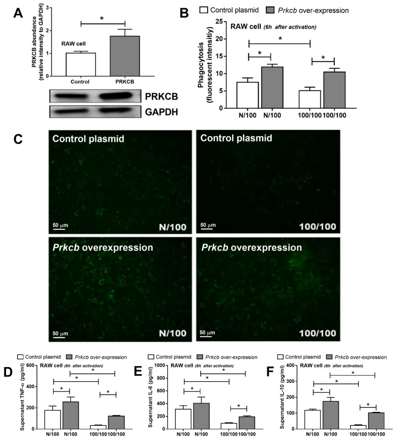
Figure 3.
The abundance of protein kinase C-β type II (PRKCB) shown by Western blot analysis from a monocyte/macrophage cell line (RAW264.7) with and without PRKCB overexpression (A) and macrophage functions after a single LPS stimulation (N/100) and sequential LPS activation (100/100; LPS-tolerance), as determined by the phagocytosis assay, (B) with representative images (green dots were phagocytosed FITC-dextran conjugated zymosan) (C) and cytokines production (D–F). Individual experiments were done in triplicate. * p < 0.05.

