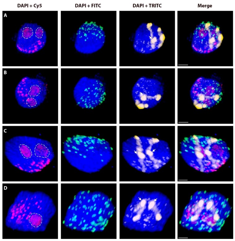
Figure 3.
Non-attachment of rye telomeres. Two nuclei ((A,B) is the same nucleus from different angles; same for (C,D)) with a pair of homologous rye del1RS.1RL chromosomes after 3D-FISH. Total genomic DNA of rye was labelled with TRITC using Nick translation (yellow colour), centromeres of both wheat and rye chromosomes were visualized using oligonucleotide probe (red colour), and telomere-specific sequence was PCR-labelled with FITC (green colour). Nuclear DNA was counterstained with DAPI (blue color). Rye telomeres without visual contact to nuclear envelope are indicated by arrows. Nucleoli are indicated by white dashed lines. Scale bar 5 µm.

