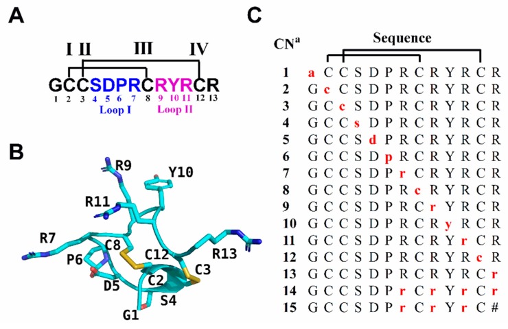
Figure 1.
Sequence and structure of RgIA and its d-amino acid scan analogues. (A) The sequence of RgIA. Disulfide connectivity of CysI-CysIII and CysII-CysIV is labeled with black lines. Amino acids in loop I and loop II are marked in blue and purple, respectively. (B) NMR structure of RgIA (PDB ID 2JUT) [30]. (C) d-amino acid scan analogues of RgIA. The amino acids indicated by red lower case letters are d-amino acids. a Compound number. The # indicates a C-terminal amide.

