
Figure 2.
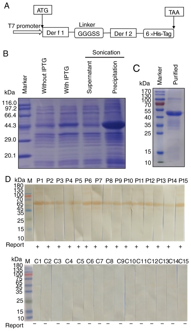
Expression, purification and IgE binding activity of rDer f 1/2. (A) Schematic diagram of the Der f 1/2 fusion protein construct. (B) SDS-PAGE of rDer f 1/2 following expression in Escherichia coli BL21(DE3) pLysS. (C) SDS-PAGE of rDer f 1/2 following purification by nickel affinity chromatography. Western blotting was conducted to determine the binding of rDer f 1/2 to specific IgE in the sera from (D) 15 house dust mite-allergic patients or (C) 15 non-allergic controls. 6×His-Tag, hexahistidine tag; Der f, allergen from Dermatophagoides farina; Ig, immunoglobulin; IPTG, isopropyl-β-D-thiogalactopyranoside; r, recombinant.