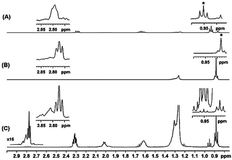
Figure 2.
(A) Selected region of a 500 MHz 1H-NMR spectrum of the lipid fraction of a lyophilized bovine milk sample in CDCl3; T, 298 K; number of scans, 256; acquisition time, 4.3 s; relaxation delay, 5 s; total experimental time ~25 min. (B) and (C) 1D TOCSY spectra with τm = 200 ms, number of scans, 256 and total experimental time ~25 min. The asterisks (*) denote the –CH3 resonances that were excited with the use of a selective shaped pulse of 80 ms.

