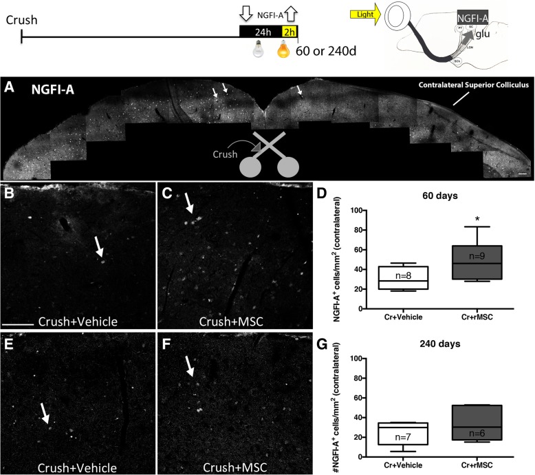
Fig. 4.
rMSC promoted synaptic reconnection in the SC. Upper panel shows the experimental design and scheme indicating that RGCs glutamatergic synapses in response to light stimulus lead to the expression of NGFI-A by post-synaptic neurons at the SC. a Photomontage of confocal images of the SC 60 d.a.c. NGFI-A+ cells (arrows) were abundant in the crushed-nerve-ipsilateral side and rare in the contralateral side. (b, c) NGFI-A+ cells in the contralateral SC 60 (b, c) and 240 (e, f) d.a.c. NGFI-A+ cells were increased in the contralateral SC of rMSC-treated animals 60 days (d) but not 240 days (g) after optic nerve crush. **P < 0.01 (Mann-Whitney test). Scale bar, 100 μm. SC: superior colliculus, PT: pretectum, LGN: lateral geniculate nucleus, SCh: suprachiasmatic nucleus, glu: glutamate

