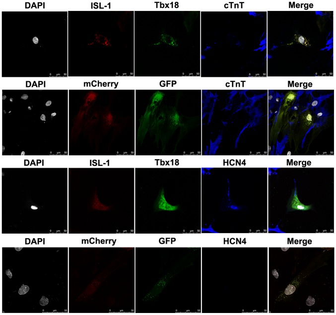
Figure 3.
Cardiac-specific proteins examined by immunofluorescence staining in differentiated ADSCs after co-culture for 7 days (magnification, ×400). Grey, nuclei stained with DAPI; red, ISL-1-ADSCs; green, Tbx18-ADSCs; and blue, representative positive staining of HCN4 and cTnT. ADSCs, adipose tissue-derived stem cells; Tbx18, T-box 18; ISL-1, insulin gene enhancer binding protein 1; HCN4, hyperpolarization-activated cyclic nucleotide-gated cation channel; cTnT, cardiac troponin T. Scale bar, 50 µm.

