Figure 4.
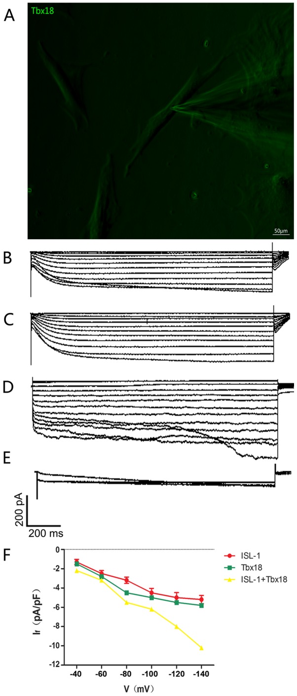
Single-cell electrophysiology of ADSCs. (A) Spindle-shaped cells were used for all electrophysiological recordings (magnification, ×200). (B) Hyperpolarization-activated inward currents recorded from ISL-1-ADSCs using the patch clamp technique. (C) Hyperpolarization-activated inward currents recorded from Tbx18-ADSCs using the patch clamp technique. (D) Hyperpolarization-activated inward currents recorded from ISL-1+Tbx18-ADSCs using the patch clamp technique. (E) Hyperpolarization-activated inward currents (If) were blocked by CsCl (4 mmol/l). (F) Current density-voltage association of ISL-1 (red, n=6), Tbx18 (green, n=6) and ISL-1+Tbx18 (yellow, n=6) groups. Tbx18, T-box 18; ISL-1, insulin gene enhancer binding protein 1; ADSCs, adipose tissue-derived stem cells; If, funny current. Scale bar, 50 µm.

