Figure 5.
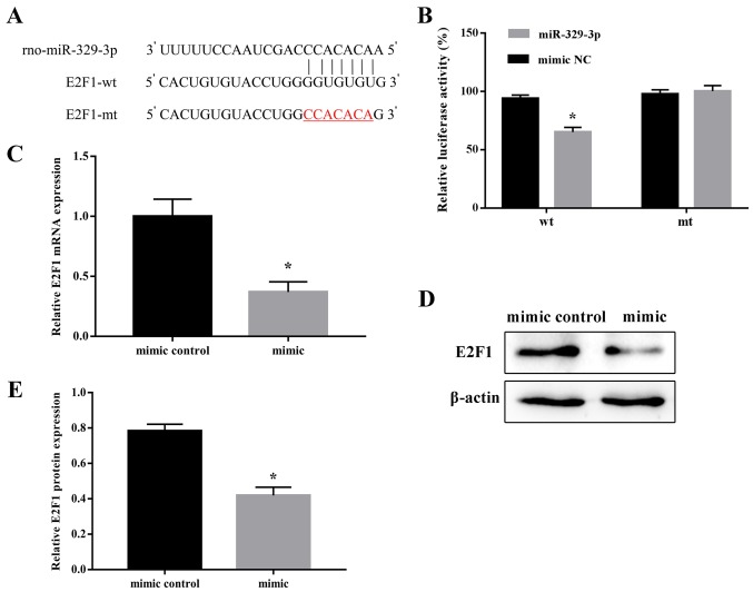
Direct targeting of E2F1 by miR-329-3p. (A) Putative miR-329-3p-binding sequence in the wt E2F1 3′-UTR. (B) Luciferase reporter assay of miR-329-3p targeting of E2F1 in 293 cells co-transfected with wt or mt E2F1 3′-UTR constructs and control or miR-329-3p mimics. (C) Reverse transcription quantitative polymerase chain reaction analysis and (D) western blot analysis of E2F1 mRNA and protein expression, respectively, in neural stem cells transfected with control or miR-329-3p mimics. GAPDH and β-actin were used as internal controls for (C) and (D) respectively. (E) Quantitative analysis of protein expression. Data are presented as the mean ± standard deviation. *P<0.05 vs. negative control mimic. E2F1, transcription factor E2F1; miR, microRNA; wt, wild-type; mt, mutated; UTR, untranslated region; rno, Rattus norvegicus.

