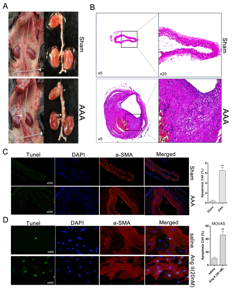
Figure 1.
AAA is induced by Ang II in ApoE−/− mice. (A) Morphology of Ang II-induced AAA and normal aortas (control) in ApoE−/− mice; white arrows indicate a typical AAA. (B) Aorta cross-sections stained with hematoxylin and eosin. (C) Representative immunofluorescence and TUNEL staining for the detection of DNA fragmentation in Ang II-induced AAA and saline-treated aortas (magnification, ×200). (D) TUNEL assay for detecting cell apoptosis in MOVAS cells treated (or not treated, saline) with Ang II by fluorescence microscopy (magnification, ×400). Light green indicates normal DNA (TUNEL-) and bright green indicates damaged DNA (TUNEL+). **P<0.01 vs. respective Sham. AAA, abdominal aortic aneurysm; Ang II, angiotensin II; ApoE−/−, apolipoprotein E-deficient; TUNEL, terminal deoxynucleotidyl transferase-mediated dUTP nick end labeling; α-SMA, α-smooth muscle actin.

