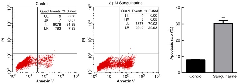
Figure 2.
Sanguinarine induces apoptosis in HLE cells. Cell apoptosis of HLE B-3 cells was detected via flow cytometry (n=3). In the control group, >90% of cells were gathered at the Q4 area, and <10% of cells underwent apoptosis (Q2+Q3 areas). However, following treatment with 2 µM sanguinarine for 24 h, cells in the Q4 area decreased to 65.7%, whereas cells in the Q2 and Q3 quadrants, which represent late and early apoptosis, respectively, were significantly increased. The results are expressed as the mean ± standard deviation. **P<0.01 vs. the control group. HLE, human lens epithelial; PI, propidium iodide.

