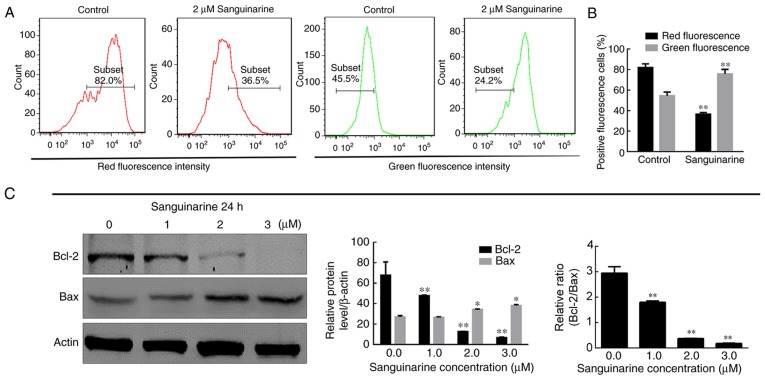
Figure 3.
MMP and HLE cell integrity are reduced following treatment with sanguinarine. (A) JC-1 dye was used as an indicator of MMP. Compared with control group, the number of red fluorescence emitting cells, which is indicative of healthy mitochondria, was markedly decreased in the sanguinarine treatment group; however, the green fluorescence emitting cells, representing damaged mitochondria, were increased. (B) Quantification of types of fluorescence-emitting HLE cells obtained following staining with JC-1 (n=3). (C) The western blotting results demonstrated that the relative proportions of Bcl-2 and Bax, key regulators of mitochondrial membrane integrity, were significantly altered following treatment with sanguinarine (n=3). The results are expressed as the mean ± standard deviation. *P<0.05 and **P<0.01 vs. respective control. HLE, human lens epithelial; JC-1, tetraethylbenzimidazolylcarbocyanine iodide dye; Bcl-2, B-cell lymphoma 2; Bax, apoptosis regulator BAX; MMP, mitochondrial membrane potential.

