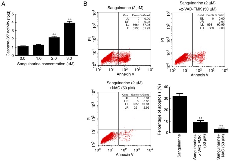
Figure 4.
Caspase and ROS serve essential roles in sanguinarine-induced apoptosis in HLE cells. (A) Caspase-3/7 activity results demonstrated that following incubation with 1, 2 or 3 µM of sanguinarine for 24 h, caspases 3/7 activity levels were significantly increased (n=3). (B) In the sanguinarine only treatment group, 31% of cells underwent apoptosis (Q2+Q3); however, in HLE B-3 cells co-treated with pan-caspase inhibitor z-VAD-FMK and sanguinarine, the apoptosis induced by sanguinarine was reduced; and cells co-treated with the ROS inhibitor NAC and sanguinarine also exhibited lower levels of apoptosis (n=3). These results suggested that ROS generation and caspase activity were required for sanguinarine-induced apoptosis. The results are expressed as the mean ± standard deviation. **P<0.01 vs. sanguinarine-only treatment. HLE, human lens epithelial; ROS, reactive oxygen species; z-VAD-FMK, benzyloxycarbonyl-Val-Ala-Asp (OMe) fluoromethylketone; NAC, N-acetylcysteine; PI, propidium iodide.

