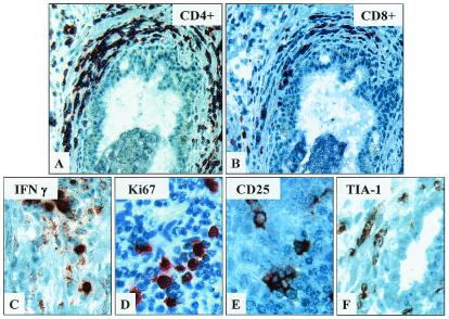
Figure 2.
T cells infiltrates within treated prostate tissues are comprised predominantly of CD4+ and a lesser amount of CD8+ T cells, including cells exhibiting markers of activation and cytotoxic potential. Immunochemical staining of CD3+ T cell-rich prostate infiltrates within treated prostate tissues demonstrates relatively greater numbers of CD4+ (A) compared with CD8+ T cells (B). Figures are at final ×400. Further staining of these sites reveals moderate numbers of cells expressing activation markers, IFN-γ (C), proliferative marker, Ki67 (D), IL2 receptor, CD25 (E), and cytotoxic granule-associated protein, TIA-1 (F). (×800.)

