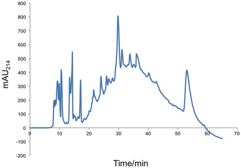
Fig. 3.
HILIC separation of 50 μg TMT-labeled peptide mixture. UV absorption at 214 nm (in mAU) is the “x” axis and elution time is the “y” axis. During the course of the chromatographic separation, a fraction is collected every 4 min, resulting in nearly 15 fractions of TMT peptide pools for subsequent total protein and phospho-peptide TiO2 enrichment analyses

