
Figure 3.
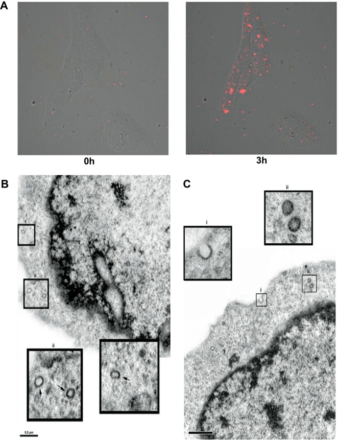
High glucose-stimulated HMC-derived exosomes can be internalized by normal HMCs. Exosomes isolated from the culture supernatants of HG treated-HMCs were labeled with fluorescent dye PHK26 and incubated with normal HMCs for 3 h. (A) Fluorescence intensity image immediately and at 3 hr after the addition of PKH26-labeled GF-Exos (red). (B) Transmission electron microscopy illustrates C-Exos incorporated by a normal HMC (arrow). Bi and Bii: Expansion of endocytosed exosomes (arrow). (C) Transmission electron microscopy showed HG-Exos incorporated by normal HMC. CI: HG-Exos being incorporated by a healthy HMC. CII: Expansion of the endocytosis of HG-Exos.