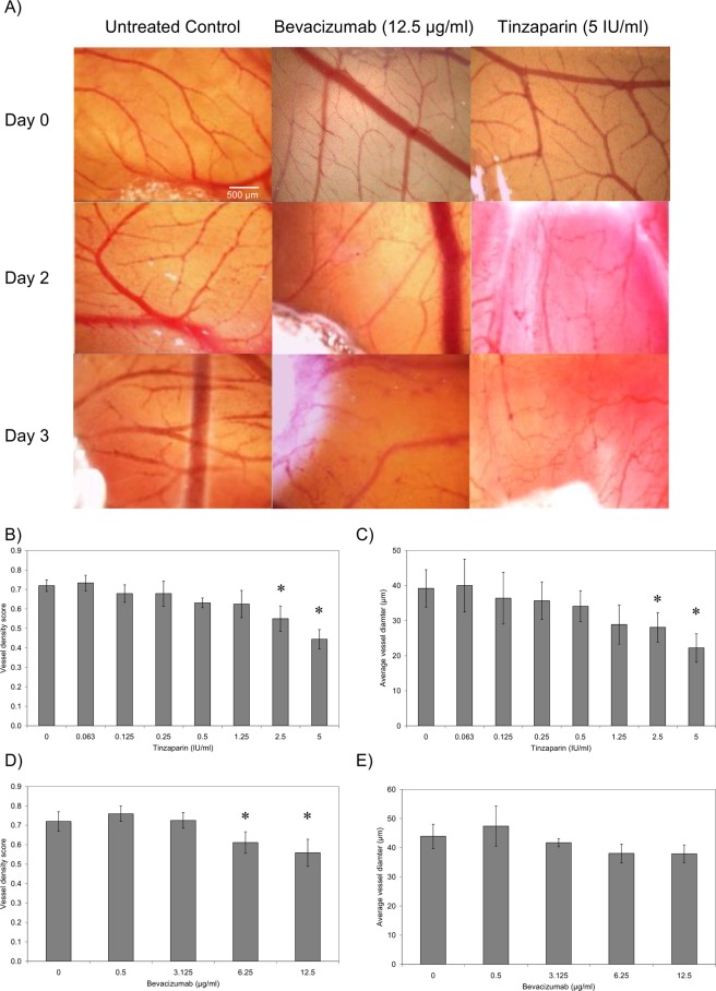
Figure 5.
The influence of Tinzaparin and Becacizumab on vascularisation in CAM. The chorioallantoic membrane assay (CAM) were prepared and examined as described in the text. Gelfoam absorbable gelatine pads were soaked with a range of concentrations of Tinzaparin (0–5 IU/ml), Bevacizumab (0–12.5 µg/ml) or PBS control which were placed on the top of the CAM. (A) The extent of vascularisation within the CAM was then examined over 3 days and Images captured using Leica Application Suite software. (B) The vessel density and (C) average vessel diameter was determined in Tinzaparin-treated CAM, and was compared to the (D) vessel density and (E) average vessel diameter in Bevacizumab-treated samples. (n = 5, *p < 0.05 vs respective PBS control).

