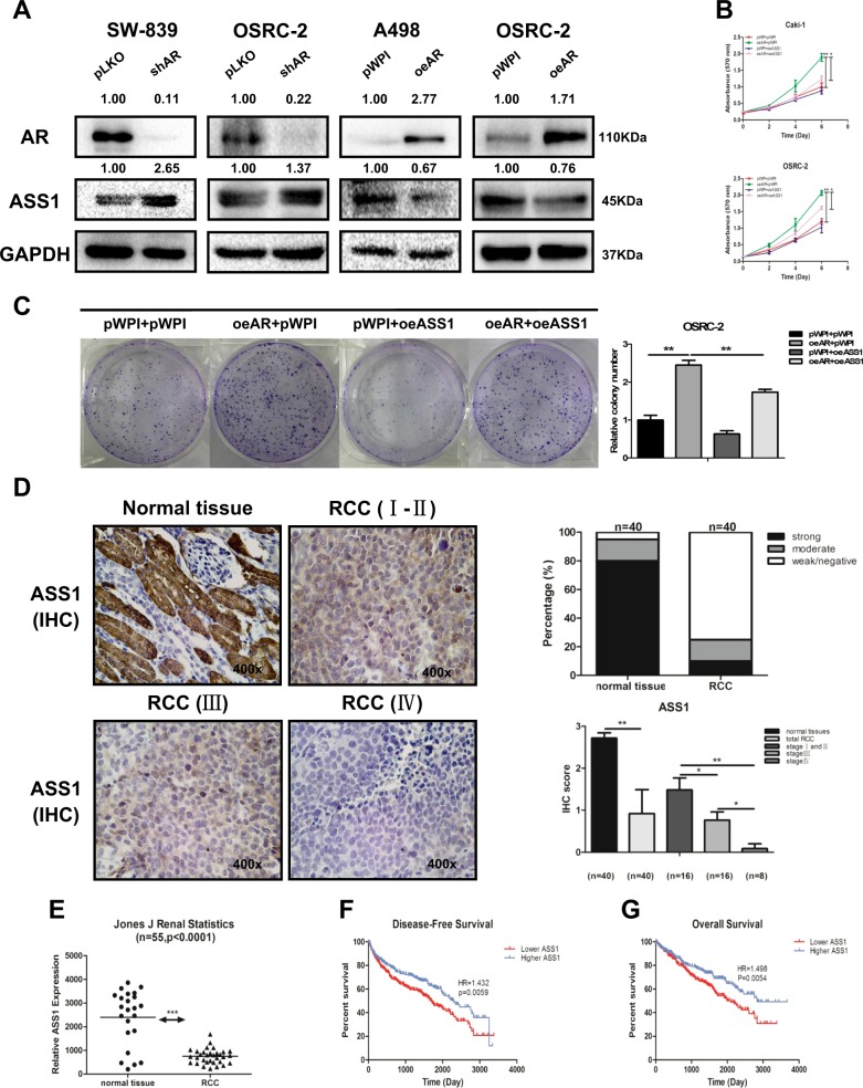
Fig. 2. AR could suppress ASS1 expression, and decreased expression of ASS1 is correlated with a worse prognosis in RCC.
a SW-839 and OSRC-2 cells were transfected with AR-shRNA or negative control. A498 and OSRC-2 cells were transfected with functional AR-cDNA and negative control. The expression of ASS1 was measured by western blot assay. b, c MTT and colony formation rescue assay reveals that AR-increased cell proliferation could be reversed/blocked after adding ASS1-cDNA in OSRC-2 and Caki-1 cells. d Immunohistochemical staining results to detect ASS1 level in 40 paired primary RCC (Stages I-IV) and adjacent normal tissues that were obtained from the Shengjing Hospital of China Medical University, Shenyang, China. Left panels show representative images, while right panels show the quantification using two different methods. Magnification is ×400. e Analysis of RCC microarray from NCBI GEO Datasets (GSE15641) shows ASS1 mRNA level in 55 RCC samples. f, g Disease-free survival curves (f) and overall survival curves (g) of RCC patients analyzed according to ASS1 expression (data were analyzed from TCGA). For (b and c), data are presented as mean ± SEM, *P < 0.05, **P < 0.01 compared to the controls

