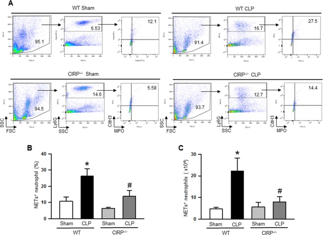
Figure 1.
Assessment of NETs in the lungs during sepsis. At 20 h of CLP or sham operation, lungs were perfused and harvested from WT or CIRP−/− mice. NET forming neutrophils in the lungs was detected by flow cytometry by staining the single cell suspension with anti-mouse Ly6G, MPO, and CitH3 Abs. (A) Representative dot plots of the frequencies of NET forming neutrophils in lungs from WT and CIRP−/− mice generated from three independent experiments are shown. Bar diagrams representing the quantitative mean values of the (B) frequencies and (C) numbers of NET forming neutrophils in the lungs are shown. Data are expressed as means ± SE (n = WT sham 4, WT CLP 6, CIRP−/− sham 6, and CIRP−/− CLP 6 mice) and compared using one-way ANOVA and SNK method (*p < 0.05 vs WT sham, #p < 0.05 vs WT CLP).

