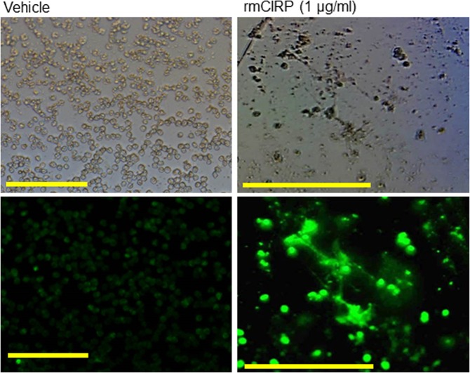
Figure 4.
Assessment of NETs in rmCIRP-treated neutrophils by florescent microscope. A total of 1 × 10⁵ BMDN grown in culture slides were treated with rmCIRP (1 µg/ml) for 4 h at 37 °C in 5% CO2. The cells were fixed with 4% paraformaldehyde and then stained with SYTOX green (2 µM) for extracellular DNA detection, followed by the assessment using fluorescence microscope (bottom panels). Web-like structures were observed in rmCIRP treated neutrophils. Top panels show the images of their counterpart under light microscope. Scale bars indicate 100 µm; original magnification ×200.

