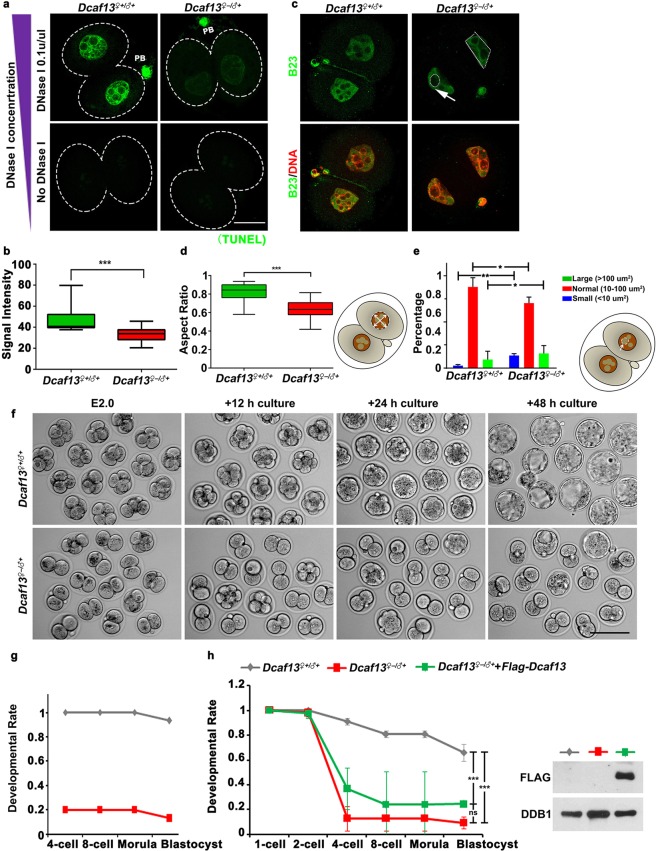
Figure 2.
DCAF13-maternally depleted embryos have abnormal chromatin tightness levels and nuclear configuration, and are arrested at the two-cell stage. (a) DCAF13-maternally depleted embryos showed decreased DNase I accessibility. Scale bar, 20 µm. (b) The positive TUNEL signal of 0.1 U/µL was indicated. *** to p < 0.001. Dcaf13♀+/♂+ (n = 12), Dcaf13♀−/♂+ (n = 20). (c) Immunofluorescence using antibodies against B23 (in green) and DNA (in red) in two-cell embryos. The abnormal nuclear morphology (white dotted line) and the abnormal nucleoli (white arrow) were indicated. Scale bar, 20 µm. Abnormal nuclear morphology was quantified at (d) Classification of nucleoli at (e). *p < 0.05, **p < 0.01. (f) Representative images for two consecutive days of in vitro culture for Dcaf13♀+/♂+ or Dcaf13♀−/♂+ embryos collected at E2. Scale bar, 100 µm. (g) Rates of embryo development of (f). (h) The numbers of zygotes, two-cell, four-cell, eight-cell, morula, and blastocysts were counted at each time point (left). Western blot results showed levels of FLAG-DCAF13 in 2-cell embryos with or without mRNA microinjection at the zygote stage. DDB1 was blotted as a loading control (right). ns to p > 0.05, *** to p < 0.001. Dcaf13♀+/♂+ (n = 64), Dcaf13♀−/♂+ (n = 71), Dcaf13♀−/♂+ with microinjection of FLAG-tagged DCAF13 (n = 57).

