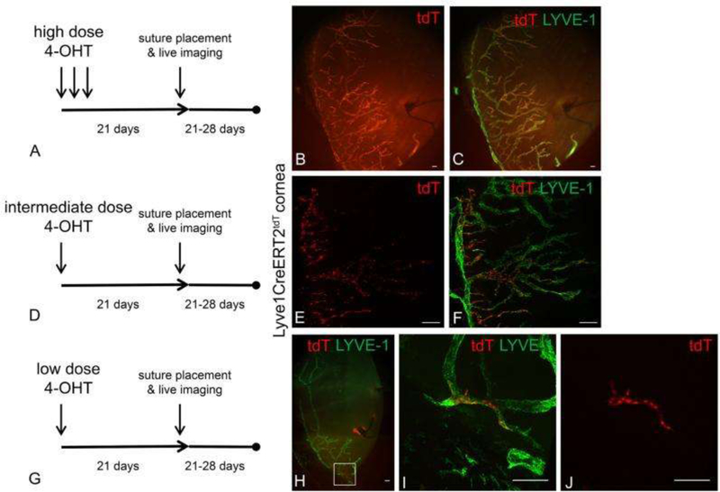
Figure 1. Tracking of the lineage of LEC progeny from tdTomato+ precursors on the background of tdTomato- lymphatic vessels in reporter mice after administration of low, intermediate, or high doses of 4-hydoxytamoxifen.
Differences in the 4-OHT dose and schedule resulted in a range of tdT expression patterns from nearly 100% to very few tdT+ LYVE-1+ LECs. After Lyve1CreERT2tdT mice were treated with high, intermediate, or low 4-OHT dosing schedules, they were rested for 3 weeks ensure the completion of tdT conversion and stability. Corneal lymphangiogenesis was stimulated using the suture-induced model of corneal inflammation. Epifluorescence microscopy revealed that after administration of high 4-OHT dosing, nearly all Lyve-1+ LEC progenies were tdT+ (A-C). After administration of the intermediate 4-OHT dosing, some of the Lyve-1+ LEC progenies were tdT+ as shown by MIP confocal microscopy (D-F). After administration of the low 4-OHT dosing, very few of the Lyve-1+ LEC progeny were tdT+ as shown by MIP confocal microscopy (G-J). In these conditions, LYVE-1+ tdT+ LEC progeny were distributed in a contiguous and relatively linear manner within a newly synthesized lymphatic vessel (J). Scale bars, 100 μm. Reprinted with permission from [46].

