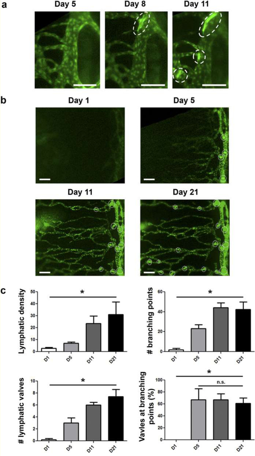
Figure 3. Intravital time course imaging showing the initiative and progressive processes of lymphangiogenesis and valvulogenesis after corneal transplantation.
(a) Initiation and progression of valve formation within limbal vessels. Scale bars, 100 μm. (b) Progressive lymphangiogenesis in parallel with valvulogenesis into the central cornea after transplantation. Scale bars, 150 μm. The vertical vessel on the right of the panel is a limbal vessel; dotted circles indicate newly formed valves. (c) Quantified data showing increase of lymphatic density, branching points, and valves over time. *P < 0.05. Reprinted with permission from [44].

