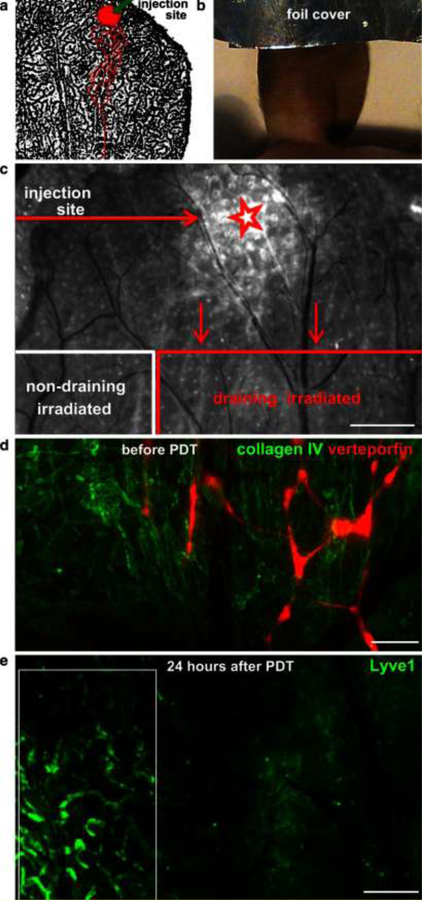
Figure 5. Fluorescence-based observation of lymphatic-specific ablation by verteporfin-PDT in the mouse ear.
(a) Schematic of the injection site and draining lymphatic vessels (red outline) over LYVE-1 staining of the mouse ear lymphatic vessels (black). (b) The injection site was covered with foil before irradiation of the rest of the ear. (c) Merged image of verteporfin fluorescence and brightfield view of the exposed dorsal ear dermis 24 h after PDT. Shown are the site of verteporfin injection (star) and the verteporfin-draining and non-draining (control) regions in the dorsal ear dermis after removal of the ventral skin and intermediate cartilage. Scale bar, 0.5 mm. (d) Fluorescence image showing verteporfin (red) draining within lymphatic vessels in the exposed dorsal skin after immunostaining for collagen IV (green). (e) Destruction of lymphatic endothelium was seen 24 h after PDT by intravital immunofluorescence staining for LYVE-1. Intact lymphatic vessels were seen in the control (non-draining) region (white box). Scale bar, 500 μm. Reprinted with permission from [52].

