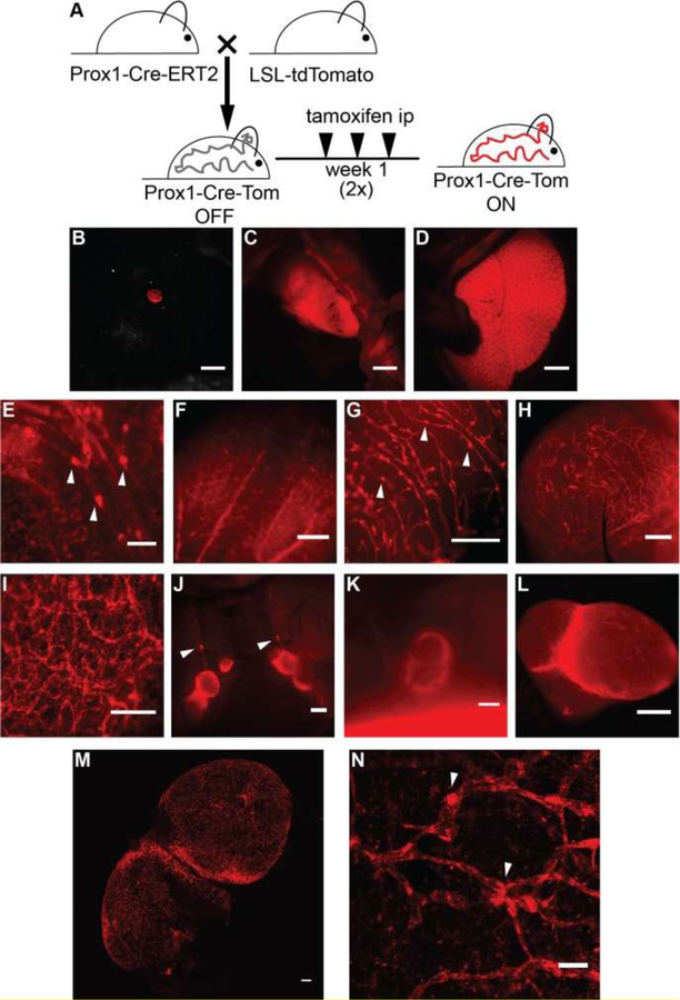
Figure 6. TdTomato expression in Prox1-positive cells upon crossing of the reporter mice with a Prox1-Cre-ERT2 line.
(A) Schematic representation of the breeding to Prox1-Cre-ERT2 mice and the tamoxifen administration regimen utilized (1 μg/g body weight in sunflower seed oil, intraperitoneally (ip), three times a week for 2 weeks). TdTomato expression was viewed under a stereomicroscope and detected in the eye (B), heart (C) and liver (D). TdTomato expression was visible in lymphatic structures in the mesentery (E), tongue (F), uterus (G), bladder (H), ear skin (ripped in half, I), lymph nodes of the neck area (J), and the inguinal lymph node (K). (L) Ex vivo image of an inguinal lymph node. (M) Confocal image of tdTomato autoflorescence showing lymphatic structures in a freshly isolated lymph node. Maximal intensity projection of a tile scan, z-stack of the lymph node is shown. (N) Confocal image (maximal intensity projection of a z-stack) of tdTomato autofluorescence showing lymphatic structures in a freshly isolated split ear sample. Arrowheads indicate lymphatic valves. Scale bars, 2000 μm (D-C), 1000 μm (J, K), 500 μm (E-I, L), and 100 μm (M-N). Reprinted with permission from [42].

