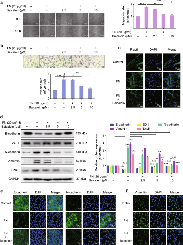
Fig. 1. Effects of baicalein on fibronectin (FN)-induced epithelial–mesenchymal transition (EMT) in breast epithelial cells.
Cells were plated on 20 μg ml−1 FN and treated with or without baicalein for 48 h. a Cell migration was measured using a wound-healing assay. Images were captured at 0 and 48 h after wounding (magnification, ×100, scale bars: 100 μm). b Cell invasion was investigated using the Matrigel-coated transwell model (magnification, ×200, scale bars: 75 μm). c Representative images from confocal microscopy analysis of F-actin organization. F-actin stained with FITC–phalloidin (green fluorescence) and cell nuclei stained with DAPI (blue fluorescence) were detected by confocal microscopy (TCS SP5; Leica, Mannheim, Germany) with Leica Application Suite Advanced Fluorescence acquisition software (original magnification ×800, scale bars: 25 μm). d Immunoblots showing expression of E-cadherin, ZO-1, N-cadherin, vimentin, and Snail in MCF-10A cells. Data represent densitometric quantification of EMT-related protein normalized with GAPDH and shown as the fold-change compared with control cells. e Representative immunofluorescence microscopy images of E-cadherin and N-cadherin after FN treatment with or without 10 μM baicalein for 48 h. Magnification, ×200, scale bars: 75 μm. f Representative confocal microscopy images of vimentin in cells after FN treatment with or without 10 μM baicalein for 48 h. Original magnification ×800, scale bars: 25 μm. Arrows indicate the network of vimentin filaments. Data are shown as mean ± SEM for three separate experiments. *P < 0.05, **P < 0.01, ***P < 0.001

