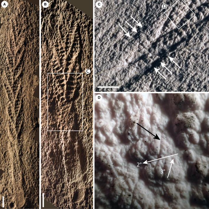
Figure 8.

Casts of specimens of Charnia masoni from Newfoundland, bed LC6. A, CAMSM X.50297.5. B, CAMSM X.50297.4. C, the basal area of the specimen in B, with second order branches visible (arrowed) on adjacent first order branches running down into the connecting region. D, rotated and furled third order branches, arrowed (black), from the specimen in B (orientation of second and third order branches indicated by white arrows). Images were retrodeformed using the constant area method. Scale bars represent 10 mm. Colour online.
