
Fig. 7.
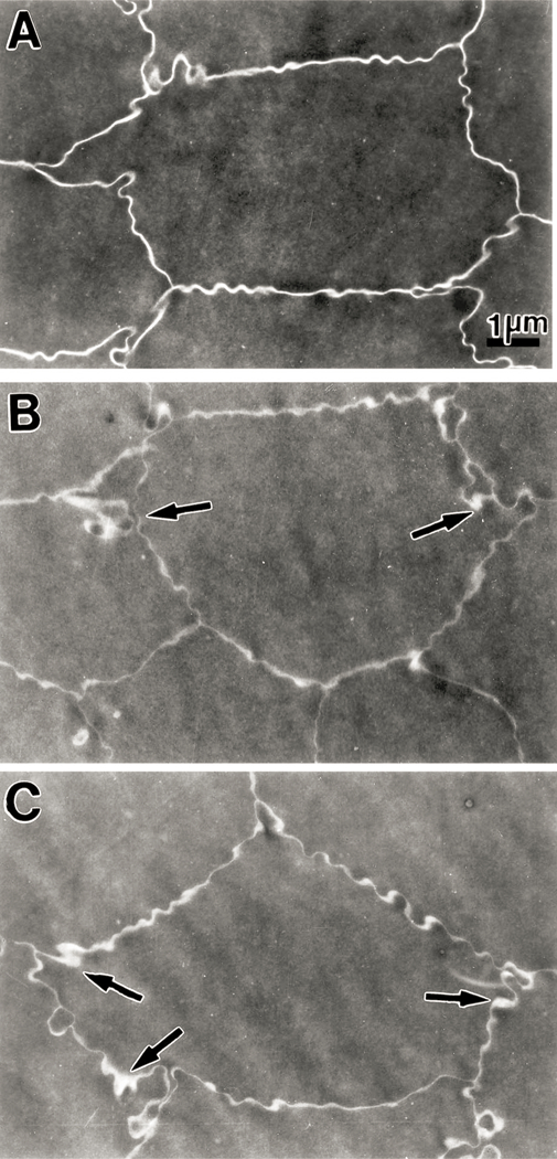
Transmission electron micrographs of the nuclear region (close to the center of the nucleus) of lenses from 23 month old guinea pigs. Micrograph A is from a section of a lens from a control animal (not exposed to UVA light) showing a uniform intercellular space between neighboring fiber cells. Micrographs B and C are from sections of a lens from an animal exposed to UVA light for five months. There is an observable distention of intercellular space between adjacent fiber cells; this is more pronounced at the Y junctions between neighboring fiber cells (arrows). Moreover, some projections and indentations between fibers are also observable at these junctions. A, B and C: × 9000.