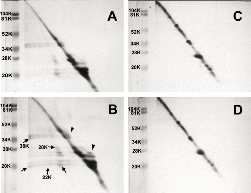
Fig. 9.

Two-dimensional diagonal SDS-PAGE patterns of urea-insoluble protein fractions of guinea pig lens nucleus and cortex. Samples of 120 µg protein were run on tube gels in the first dimension without prior treatment with mercaptoethanol. The tube gels were incubated for 30 min in buffer containing mercaptoethanol and applied on the second dimension. The presence of off-diagonal spots (arrows) indicates disulfide-crosslinked proteins. Also note the protein which appears to the right of the diagonal (arrowheads). (A) Lens nucleus from a 23 month old control animal. (B) Lens nucleus from a 23 month old animal after five months of exposure to UVA light. (C) Lens cortex from a 23 month old control animal. (D) Lens cortex from a 23 month old animal after five months of exposure to UVA light.
