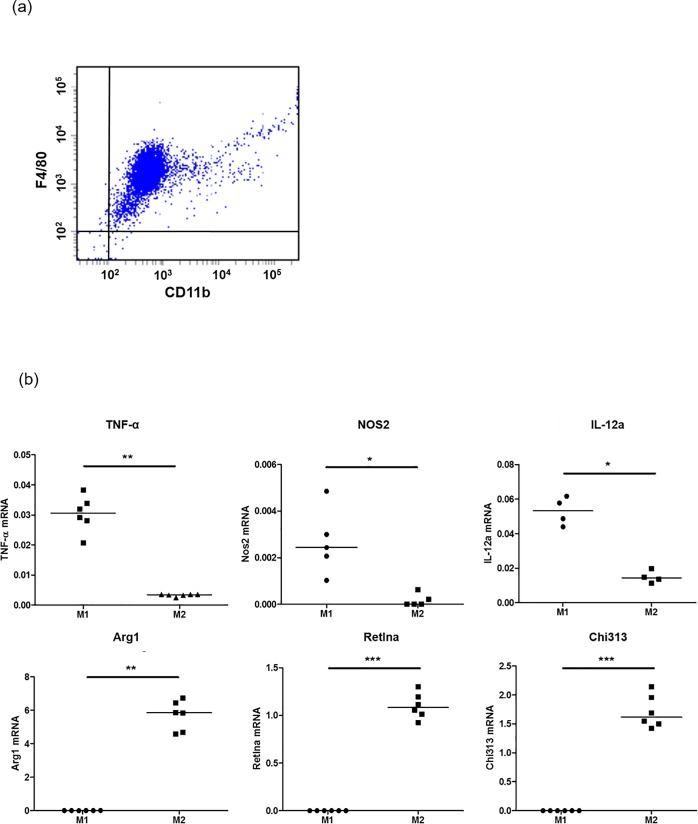
Fig 1. Identification of M1 and M2 macrophages.
Panel (a). Murine bone marrow-derived macrophages were analyzed by flow cytometry using antibodies against F4/80 and CD11b. F4/80+/CD11b+ cells are analyzed as macrophages. Panel (b). Using quantitative RT-PCR, M1 macrophages were identified by expression of Tnf, Nos2, and IL-12a, t and M2 macrophages were identified by expression of Arg1, Retnla, and Chi313 Mann-Whitney U-test; *P < 0.05, **P < 0.001, ***P < 0.0001.

