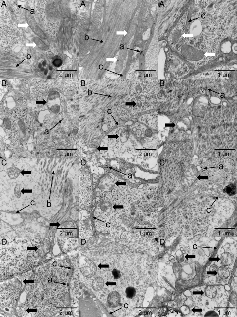
Fig 7. Transmission electron microscope images of the foot muscle of Ruditapes philippinarum.
(A), (B), (C) and (D) represent the DO groups of 6.0, 2.0, 1.0 and 0.5 mg L-1, respectively. White arrows point to normal mitochondria while black arrows point to mitochondria with collapsed cristae (judged by sparse cristae) and vacuolization (judged by shallower staining). Cell vacuolization was apparent according to the shallower cell-staining. Some other cellular structures for point of reference are listed as follows: ‘a’ refer to glycogen granules; ‘b’ refer to myofilaments; ‘c’ refer to cell membrane.

