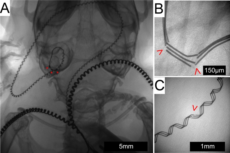
Fig 8. In-vivo x-ray imaging.
X-ray imaging shows an intracochlear position of the array in the left cochlea of a live subject with the electrode contacts labeled by a red “*” (A). The lead wire can be seen traveling across the posterior base of the skull to meet the 2 extracochlear electrode leads as they head toward the connector located on the back (not seen in this image). Fractures (red arrowhead) occurred in both the straight (B) and helixed portions (C) of the array lead wire.

